TL;DR
TMS therapy and neurofeedback are both non-invasive approaches that support brain health, but they work in different ways. TMS uses targeted magnetic stimulation, while neurofeedback trains brain activity over time. The right choice depends on your symptoms, goals, and care plan.
If you're exploring options to support brain health, you may be asking: What's the difference between TMS therapy vs neurofeedback, and which one is right for me?
Both approaches are increasingly used to support conditions related to mood, cognition, and neurological function. But while they may sound similar, they work very differently.
In this guide, we'll break down how TMS therapy and neurofeedback compare, what each is designed to do, and how to choose the most appropriate path based on your needs.
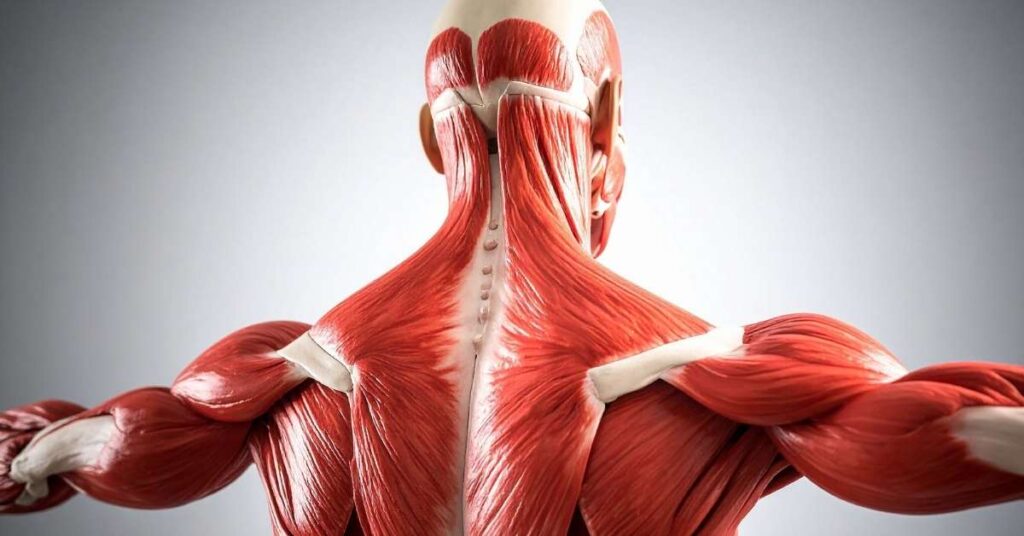
TMS therapy is a non-invasive treatment that uses magnetic pulses to stimulate targeted areas of the brain.
Transcranial Magnetic Stimulation (TMS) focuses on specific regions associated with mood regulation and cognitive function. Delivering controlled magnetic pulses, it helps activate underperforming neural pathways.
TMS is FDA-cleared for multiple conditions, including major depressive disorder, obsessive-compulsive disorder, and nicotine dependence.
It has been in clinical use since 2008 and is now widely available with established treatment protocols.
Standard TMS treatment typically involves 20-30 sessions over 4-6 weeks, with each session lasting 30-40 minutes, delivered 5 days per week.
TMS is commonly considered when:
Learn more about how non-invasive TMS treatment works and whether it may fit into your care plan.

Neurofeedback is a brain-training approach that uses real-time monitoring of brain activity via EEG to provide feedback intended to help regulate brainwave patterns.
This method is often described as "exercise for the brain."
Neurofeedback may support:
However, current research shows limited and inconsistent results. For conditions like ADHD, high-quality studies using blinded assessments show no significant benefit compared to control conditions.
For depression, neurofeedback is considered "possibly efficacious" (level 2/5 evidence), with treatment efficacy only significant in randomized controlled trials when accounting for experimental design.
Methodological concerns and study quality issues limit the strength of these findings.
Some findings suggest that observed improvements may be related to nonspecific effects, such as engagement or placebo response, rather than direct changes in brainwave activity.
For a deeper look, explore neurofeedback therapy services and how this approach supports long-term brain function.
The main difference between TMS therapy vs neurofeedback is how they influence brain activity. TMS stimulates, while neurofeedback attempts to train.
Both approaches benefit from detailed assessment. Services like cognitive testing and evaluation, or cognitive function testing, help guide more precise care decisions.
Choosing between TMS therapy and neurofeedback starts with a comprehensive evaluation of your brain health and goals.
A care team may consider:
From there, a personalized plan may include:
The focus is always on evidence-based care, tailored to your needs.
Understanding your options early allows for more effective, personalized care.
When you take time to explore approaches like TMS therapy vs neurofeedback:
Most importantly, it empowers you to take an active role in your neurological health.

If symptoms are affecting your daily life or not improving, it may be time to explore advanced, non-invasive options.
Consider seeking guidance if:
Professional evaluation helps clarify which path, TMS therapy, neurofeedback, or both, may best support your goals.
Neither is universally better. TMS has strong clinical evidence and FDA clearance for specific conditions such as depression, OCD, and nicotine dependence.
Neurofeedback has limited evidence and is not supported as a stand-alone treatment for most conditions.
The best option depends on your individual needs.
This combination is being explored, but current evidence is insufficient for clinical recommendations due to small sample sizes, short follow-up periods, and potential publication bias.
TMS has been FDA-cleared since 2008 and is widely available in many clinical settings.
However, access can still vary depending on location, provider availability, and awareness.
While neurofeedback has been explored in various research settings, there is no established evidence of routine clinical use by NASA or similar organizations.
Choosing between TMS therapy and neurofeedback doesn’t have to be overwhelming.
Here’s a simple recap:
Book a strategy call with Universal Neurological Care to explore your options.
Loss of muscle mass is often blamed on aging, but in some cases, the real issue involves neurological causes of muscle weakness that disrupt how nerves communicate with muscle tissue.
When that signal pathway breaks down, strength fades, muscles shrink, and everyday movements become unreliable.
One leg may look smaller than the other. Grip strength may decline without explanation. Fatigue sets in faster than it used to.
Not all muscle loss is due to inactivity. Sometimes the nervous system is involved, and knowing the difference can change what happens next.
Let’s walk through when muscle weakness may be neurological, what conditions can cause it, and how it’s evaluated.
Loss of muscle mass, also called muscle atrophy, means muscle tissue becomes thinner and weaker. It can develop slowly from inactivity, poor nutrition, or aging. That’s called disuse atrophy.
Neurogenic atrophy is different. It happens when nerves fail to properly stimulate the muscle. Without regular nerve input, muscle fibers shrink. The process can move faster and may affect one side of the body more than the other.
That uneven pattern is often the first clue that something neurological may be happening.
Muscle movement depends on a chain of communication.
The brain sends signals down the spinal cord. Those signals travel through peripheral nerves. At the neuromuscular junction, the nerve releases chemical messengers that trigger muscle contraction.
If any part of that pathway is disrupted, it can be the brain, spinal cord, nerve root, peripheral nerve, neuromuscular junction, or muscle fiber, and weakness follows.
Over time, reduced activation leads to visible loss of muscle mass.
The pattern of weakness often reveals where the breakdown is occurring.
Different conditions affect different parts of the system. The symptoms may overlap, but the underlying cause can vary significantly.
Peripheral neuropathy affects the nerves outside the brain and spinal cord. Diabetes is one of the most common causes. Autoimmune disorders, toxin exposure, infections, and inherited conditions like Charcot-Marie-Tooth disease can also contribute.
Symptoms often begin in the feet or hands and may include numbness, tingling, burning pain, and weakness. Over time, muscles in those areas may shrink due to reduced nerve signaling.
Motor neuron diseases such as amyotrophic lateral sclerosis (ALS) and spinal muscular atrophy affect the nerve cells responsible for voluntary movement.
When motor neurons deteriorate, muscles lose direct input. Weakness progresses. Muscle wasting becomes more noticeable. Twitching or cramping may occur as the remaining motor units struggle to compensate.
A herniated disc or spinal degeneration can compress a nerve root. If that nerve carries motor fibers, the muscle it supplies may weaken.
This often presents as hand pain, sometimes in a specific region, for example, the calf, shoulder, or calf pain, and sometimes accompanied by radiating pain. If compression continues, loss of muscle mass can develop in that distribution.
The neuromuscular junction is the point where nerve and muscle communicate. Conditions such as myasthenia gravis interfere with that communication.
Weakness may fluctuate and worsen with repeated use. Drooping eyelids, difficulty swallowing, and speech changes can appear early. Muscle wasting may develop over time if weakness persists.
Some conditions affect the muscle fibers directly rather than the nerves that control them. Muscular dystrophy is one of the clearest examples.
According to Johns Hopkins Medicine, muscular dystrophy refers to a group of inherited diseases that cause progressive muscle weakness and muscle tissue wasting. Different types vary in age of onset, the muscles involved, and the rate of progression.
Duchenne muscular dystrophy, the most common form, typically begins in early childhood and primarily affects boys.
Becker muscular dystrophy follows a similar pattern but progresses more slowly.
Other forms, including limb-girdle, facioscapulohumeral, myotonic, and oculopharyngeal muscular dystrophy, affect different muscle groups and may appear later in life.
These conditions demonstrate how genetic and neuromuscular disorders can directly impair muscle structure, leading to progressive loss of muscle mass over time.
Inflammatory muscle diseases such as polymyositis and dermatomyositis also fall into this category. In these cases, the immune system attacks muscle tissue, leading to weakness and gradual atrophy.

Evaluation begins with a detailed neurological examination assessing strength, reflexes, coordination, and sensation.
If nerve involvement is suspected, diagnostic testing helps clarify the site of the disruption. Electromyography (EMG) and nerve conduction studies (NCV) measure how well electrical signals travel from nerve to muscle and can distinguish between nerve-related and primary muscle causes of weakness.
Additional testing may include blood tests to screen for autoimmune or metabolic disorders, as well as brain or spine imaging if structural causes are suspected.
In more complex cases, a neuromuscular evaluation may also involve autonomic testing, cognitive assessment, or functional balance testing to determine whether broader nervous system involvement is contributing to symptoms.
Identifying the exact source of weakness allows treatment to be tailored rather than generalized.
If loss of muscle mass is happening without a clear reason, especially if it’s uneven, progressive, or paired with numbness, twitching, or balance changes. Don’t wait it out. Muscle weakness tied to nerve dysfunction tends to worsen when the cause isn’t identified early.
At Universal Neurological Care, evaluation goes beyond a basic strength check. Neuromuscular assessment may include electromyography (EMG/NCV) testing to assess nerve-muscle communication, along with targeted neurological exams and additional diagnostic studies when needed.
Yes. When nerves fail to properly stimulate a muscle, the muscle can shrink over time. This is called neurogenic atrophy and can progress if the underlying nerve issue isn’t addressed.
It depends on the cause. Muscle loss from temporary nerve compression may improve with treatment. Progressive nerve diseases may require long-term management rather than reversal.
Doctors use a neurological exam, along with tests such as EMG and nerve conduction studies, to determine whether weakness is due to nerve dysfunction or to the muscle itself.
Asking what sarcopenia is usually isn’t something people ask until their body starts feeling different. Your strength may drop off. The stairs can feel harder. Trying to get up from a low chair takes a second try.
Some muscle loss happens with age. Sarcopenia is when that loss moves past “normal” and starts affecting balance, mobility, and daily life.
Doctors now classify it as a disease because of how strongly it’s linked to falls, fractures, hospital stays, and slower recovery from illness or surgery.
If you’ve noticed strength slipping, or you’re caring for someone who has, it helps to know what’s going on. Here’s what causes sarcopenia, how it’s diagnosed, and what actually helps.
Sarcopenia is the gradual loss of muscle mass, strength, and physical performance that occurs with aging. Everyone loses some muscle mass over time, but sarcopenia describes a decline that begins to affect daily function.
It’s now classified as a medical condition because of how strongly it’s linked to falls, fractures, hospital stays, and slower recovery from illness or surgery. The diagnosis typically involves three things: reduced muscle strength, reduced muscle mass, and slower physical performance.
It most often affects adults over 60, especially those who are sedentary or living with chronic health conditions.
Sarcopenia doesn’t happen for just one reason. Muscle loss builds slowly over time, and usually more than one factor is involved. Age sets the stage, but biology, lifestyle, and overall health all influence how quickly strength declines.

Getting older changes how muscles behave. Hormone levels drop, including testosterone and growth-related hormones that help maintain muscle tissue.
Nerve signals that tell muscles to contract become less efficient. The body also becomes less responsive to dietary protein, meaning muscle repair after meals or exercise isn’t as strong as it once was.
Fast-twitch muscle fibers, the ones that generate power and help prevent falls, shrink more than endurance fibers. That’s often why strength fades before muscle size visibly changes.
Muscles need regular challenge. When daily movement decreases, muscle mass declines more quickly. Long stretches of sitting, limited resistance training, or reduced overall activity can all accelerate the process.
Even moderate activity helps slow the decline, but complete inactivity accelerates it.
Protein provides the building blocks for muscle repair. Many older adults eat less protein than their bodies need. Without enough dietary support, rebuilding muscle becomes difficult, even with exercise.
Over time, that imbalance contributes to a gradual loss.
Several chronic conditions increase the risk of sarcopenia, including diabetes, chronic kidney disease, heart failure, COPD, and cancer.
Some affect muscle metabolism directly. Others reduce energy levels or mobility, which, in turn, contributes to muscle decline.
Low-grade inflammation, which tends to increase with age, can also promote muscle breakdown. That steady inflammatory state makes it harder for the body to preserve lean tissue.
Most people with sarcopenia don’t have just one cause. Aging, inactivity, metabolic changes, and chronic disease often overlap and reinforce one another.
Sarcopenia develops gradually, which makes it easy to miss early on. The common signs include:
As it progresses, everyday tasks may require more effort. Carrying groceries, getting out of a car, or walking longer distances can become challenging.
Sarcopenia doesn’t respond to a quick fix. No medication reverses it.
Treatment focuses on rebuilding strength and slowing further loss through consistent, targeted action. What works is practical and proven.
Strength training is the foundation of treatment. Muscles respond to load, even later in life. Lifting weights, using resistance bands, or performing controlled bodyweight exercises two to three times per week can improve muscle strength and physical performance.
You don’t need to do extreme workouts. It’s steady, progressive resistance that challenges the muscle enough to trigger adaptation. Even adults in their 70s and 80s can regain measurable strength when training is consistent.
Muscle repair depends on adequate protein. Many older adults unintentionally under-consume it.
According to the Journal of the American Medical Directors Association, most clinical guidelines recommend 1.0-1.2 grams of protein per kilogram of body weight per day, often distributed across 20–35 grams per meal. Spreading intake evenly across meals improves muscle protein synthesis.
Exercise without protein limits recovery. Protein without resistance training does little on its own. Together, they reinforce each other.
Chronic diseases that limit mobility or affect metabolism should be addressed directly. Poorly controlled diabetes, chronic inflammation, and hormonal imbalances can all accelerate muscle decline.
If weakness appears sudden, asymmetric, or more severe than expected for age, further evaluation may be needed to rule out neuromuscular disease.

Muscle weakness isn’t always just “getting older.” When strength drops faster than expected, balance becomes unstable, or falls become more frequent, it’s worth taking a closer look.
At Universal Neurological Care, evaluation may include neuromuscular assessment and diagnostic testing, such as EMG/NCV studies, to determine whether nerve dysfunction is contributing to weakness.
For patients experiencing balance issues, additional functional testing can help clarify what’s driving the decline.
If muscle loss has begun to affect mobility or daily activities, a structured rehabilitation plan may help restore function and reduce the risk.
Muscle loss can start as early as your 30s, but it typically becomes more noticeable after age 60. The rate of decline increases with each decade, especially without regular strength training.
Everyone loses some muscle with age. Sarcopenia refers to a more significant decline that affects strength and physical performance, not just muscle size. It starts to interfere with daily tasks like walking, climbing stairs, or getting up from a chair.
When people look into the benefits of alpha stimulation therapy, they’re often trying to find something that supports the nervous system without adding another medication.
Anxiety, chronic stress, sleep disruption, and mild mood imbalance frequently stem from patterns of overactivation in the brain.
Alpha Stimulation Therapy is a non-invasive brain stimulation option and is often discussed alongside TMS therapy within comprehensive mental health care programs.
Both approaches fall under the umbrella of neuromodulation, but they differ in intensity, delivery, and clinical use. Knowing where Alpha Stimulation Therapy fits helps set realistic expectations.

Alpha Stimulation Therapy uses cranial electrotherapy stimulation (CES). Small electrodes attach to the earlobes and deliver a very low microcurrent.
The intensity is subtle and is designed to influence neural signaling related to arousal and mood regulation.
It aims to gently influence stress-related circuits and support more stable patterns of activity over time. Sessions are typically brief and can be performed at home under medical guidance.
CES is not the same as electroconvulsive therapy. It does not require anesthesia, does not induce seizures, and uses a much lower level of current.
One clear advantage is that it is medication-free. For adults who are sensitive to side effects or prefer adjunctive options, that matters.
Clinical reports and smaller trials suggest it may:
Another benefit is tolerability. Reported side effects tend to be mild and uncommon, such as temporary skin irritation or headache.
Larger clinical trials have examined Alpha-Stim devices more closely. A multicenter randomized controlled trial published in The Lancet Psychiatry in 2023 (Morriss et al.) evaluated Alpha-Stim AID in adults with major depression.
The study found the device to be safe and well-tolerated, though it did not demonstrate superiority over sham treatment for reducing depressive symptoms.
Findings like this highlight an important point: outcomes depend on diagnosis, severity, and how the therapy is integrated into care.
In anxiety-focused use, earlier research and clinical practice reports suggest more favorable symptom improvement, though results can vary.
Alpha Stimulation Therapy delivers low-intensity microcurrent and is generally used daily at home.
TMS therapy, by contrast, uses magnetic pulses applied in a clinical setting and is commonly recommended for treatment-resistant depression.
The difference reflects intensity and clinical indication. Alpha stimulation may be considered for anxiety, stress, and sleep-related concerns.
TMS therapy is often reserved for more persistent or severe depressive conditions.
Deciding between them depends on symptom severity, history of treatment response, and overall mental health goals.
The answer depends on how the symptoms present. Anxiety driven by chronic stress and disrupted sleep may require a different strategy than moderate to severe depression that has not improved with medication.
A structured clinical evaluation helps clarify whether Alpha Stimulation Therapy makes sense on its own or as part of a broader neuromodulation plan.

If anxiety, stress, or sleep problems continue to affect daily life, the next step is a careful review of symptoms and treatment history.
Understanding how your nervous system is functioning helps determine whether Alpha Stimulation Therapy or another neuromodulation approach is appropriate.
At Universal Neurological Care, treatment decisions are based on clinical evaluation rather than device preference.
Neuromodulation services, including Alpha Stimulation Therapy and TMS therapy, are considered within a comprehensive mental health plan tailored to individual needs.
Clinical research indicates it is generally well-tolerated. The reported side effects are uncommon and usually mild, such as temporary skin irritation or headache.
In the United States, Alpha-Stim devices are FDA-cleared for anxiety, insomnia, and pain. Clearance for depression differs by country and indication.
Some individuals notice changes in sleep or physical tension within a few weeks of consistent use. Response time varies depending on the condition and severity.
TL;DR
If neuropathy symptoms such as numbness, burning pain, weakness, or digestive changes are persistent, worsening, or affecting daily life, it may be time to see a neurologist for specialized evaluation and support.
Many people living with tingling, numbness, or nerve pain eventually ask the same question: when to see a neurologist for neuropathy. Because neuropathy can progress gradually and present differently for each person, it’s not always clear when general care is no longer enough.
This article outlines the most common signs that specialist neurological care may be beneficial, explains how neurologists support people with neuropathy, and helps patients and caregivers understand when informed next steps matter most.
Neuropathy refers to damage or dysfunction of nerves that transmit signals between the brain, spinal cord, and body.
Most commonly, neuropathy affects peripheral nerves in the hands and feet, but it can also involve muscles, balance systems, and autonomic functions such as digestion or circulation. Because nerves play many roles, symptoms can vary widely in intensity and presentation.
Understanding neuropathy early helps individuals and caregivers make informed decisions about neurological care and support pathways.

Neuropathy symptoms may involve sensory, motor, or autonomic nerves.
Common symptoms include:
Potential causes or contributing factors may include:
Because symptoms may overlap with other conditions, neurological expertise is often key to clarity.
It may be time to see a neurologist when neuropathy symptoms are no longer stable, manageable, or clearly understood.
Signs that specialist care may be appropriate include:
According to clinical diagnostic algorithms synthesized by OpenEvidence, consultation with a neurologist or neuromuscular specialist is specifically indicated when neuropathy presents with atypical features.
These include asymmetrical symptoms, an acute or subacute onset, a rapidly progressive course, motor predominance, or prominent autonomic involvement. Recognizing these specific signs ensures that patients can access specialized evaluations, such as electrodiagnostic studies or nerve biopsies, that are crucial for diagnosing and managing complex cases.
Neurologists specialize in evaluating nerve pathways and can help determine whether symptoms involve peripheral nerves, central nervous system processes, or related conditions. They may connect you with appropriate diagnostic services, like Diagnostic Procedures, and develop a tailored care plan.

Seeing a neurologist provides focused insight and coordinated neurological care.
Key benefits may include the following:
This patient-focused approach prioritizes understanding, function, and quality of life rather than quick assumptions.
Neuropathy evaluation focuses on assessing nerve health and overall neurological function.
A neurologist may:
Management is individualized, focusing on symptom relief, functional support, and informed long-term planning without guarantees or alarmist language. Neurologists may also explore related support options such as TBI Rehabilitation if symptoms intersect with balance or injury recovery.
Early neurological guidance helps reduce uncertainty and unnecessary delays.
Benefits of early support include:
Even when symptoms feel mild, clarity can provide reassurance and direction.
Yes. A neurologist evaluates nerve function, identifies contributing factors, and helps guide supportive care and long-term neurological management.
In some cases, neuropathy may be associated with swelling, particularly if circulation, inflammation, or autonomic nerve involvement is present. Evaluation helps clarify the cause.
Yes. Certain forms of neuropathy can affect autonomic nerves that regulate digestion, leading to diarrhea or other bowel changes.
Progressive numbness, persistent nerve pain, muscle weakness, balance issues, or unexplained sensory or digestive changes are common reasons to seek neurological guidance.
Book a strategy call with Universal Neurological Care to discuss symptoms and care pathways.
When people search for neurofeedback vs TMS for anxiety, they’re usually tired of trying things that haven’t worked.
Anxiety can lock your body into a constant alert state with a tight chest, racing thoughts, and poor sleep, and no amount of willpower fixes that.
The issue often lies in how the brain regulates stress. Neurofeedback and TMS both work at that level, but they do it in very different ways.
Knowing the difference matters because the right fit depends on how your anxiety shows up day to day.
Neurofeedback works by helping the brain recognize when it’s stuck in an alert state and gently guiding it back toward steadier patterns.
Sensors track brain activity in real time, and the feedback gives the brain a signal when it moves in a calmer direction. Over repeated sessions, the brain starts to remember how to settle on its own.
Changes usually arrive quietly. Sleep tends to improve first. The body feels less jumpy during the day. That constant sense of tension or restlessness eases before anxious thoughts fully slow down.
Clinically, that order makes sense. When the nervous system stays on high alert, the mind follows. Once the body settles, the thoughts often become easier to manage.
That’s why neurofeedback often fits anxiety that comes with poor sleep, panic symptoms, migraines, post-concussion issues, or a constant sense of physical tension.
The goal isn’t to force the brain into a new state. Instead, it’s about helping it relearn what a regulated state feels like and return there more easily.
Research supports this pattern. In a clinical study by Kosari and colleagues, patients receiving neurofeedback showed improved sleep quality over time, with a noticeable reduction in the time to fall asleep.
Sleep improvements emerged early and persisted at follow-up, suggesting that regulation often begins with rest before spreading to other symptoms.

TMS approaches anxiety from a different angle. Rather than training the brain from within, it applies magnetic stimulation from the outside.
Specific brain areas involved in mood and control receive repeated pulses while the patient remains passive during sessions.
This approach can be helpful when anxiety is closely tied to depression or when symptoms feel heavy and unresponsive.
Some people notice changes faster than they would with training-based methods, which can matter when anxiety feels stuck or layered with low mood.
People looking into TMS therapy in Jacksonville often ask whether TMS treats anxiety directly. The answer usually depends on what’s driving the anxiety.
When depression, reduced mental control, or emotional blunting plays a major role, stimulation-based care can be appropriate.
The question usually isn’t which option is better. It’s whichever one fits the pattern of symptoms.
Neurofeedback focuses on regulation and tends to help anxiety linked to hyperarousal, sleep disruption, and physical tension.
TMS focuses on stimulation and may suit anxiety tied to low mood or difficulty engaging mental control.
Neurofeedback often leads to lasting changes because the brain learns a new baseline. TMS can lead to quicker shifts, though some people may need follow-up sessions to maintain results.
When the approach doesn’t match the underlying issue, progress can stall even if the treatment itself is sound.

Sometimes both approaches are appropriate, but the sequence matters. One method may reduce symptom intensity, while the other supports longer-term stability.
That decision typically depends on sleep patterns, symptom history, and early nervous system responses. Careful evaluation tends to lead to more consistent outcomes than trial-and-error treatment.
If anxiety continues to affect sleep, focus, or daily function, the next step is a clinical evaluation that looks at how your nervous system is actually operating.
A specialist can help identify whether symptoms point toward dysregulation, mood-related changes, or a combination of factors, which makes choosing between neurofeedback, TMS, or a coordinated approach far more precise.
At Universal Neurological Care, evaluations focus on sleep patterns, symptom history, and neurological function before recommending treatment.
Care decisions stay guided by objective findings and clinical experience, with the goal of steady improvement that holds up over time.
TMS can lead to earlier changes for some people, especially when symptoms feel heavy or resistant. Neurofeedback usually works more gradually, with sleep and physical calm improving before anxious thoughts settle.
Most people tolerate both well, but responses differ. A poor match or poorly adjusted protocols can increase discomfort, which is why early oversight matters.
The choice often depends on sleep patterns, symptom history, and early nervous system response. Anxiety driven by constant alertness tends to respond differently than anxiety tied to low mood. A clinical review helps clarify the starting point.
The benefits of neurofeedback therapy interest many people who want a more brain-based, non-drug way to improve focus, mood, or stress.
At the same time, it is not magic, and science is still evolving.
Neurofeedback is a real, EEG-based method with decades of research behind it; however, the science is still evolving, and results vary from person to person.
Used effectively, it can help some patients regulate brain activity more efficiently and derive greater benefits from standard treatments, such as medication and psychotherapy.
According to the National Library of Medicine, neurofeedback is a form of EEG biofeedback. Small sensors on your scalp measure brainwaves while you sit in front of a screen.
A computer converts those signals into real-time feedback:
When your brain produces more regulated patterns (for example, calmer or more focused activity), the system “rewards” it by making the screen or sound more pleasant.
When it drifts away, the reward fades. Over many repetitions, your brain learns what a better-regulated state feels like and becomes more practiced at achieving it on its own.
Neurofeedback has been studied across many conditions. Evidence quality varies, but several benefits stand out in day-to-day clinical use.
Many people with anxiety live in a constant “on edge” state. Neurofeedback can target overly fast or unstable brainwave patterns associated with hyperarousal and help establish more relaxed rhythms.
Commonly reported changes:
It does not eliminate normal worry, but it can lower the baseline, allowing coping skills and therapy to work more effectively.
Depression often involves sluggish activity in some networks and overactive rumination in others. Neurofeedback aims to rebalance these patterns and improve functions such as motivation, planning, and cognitive flexibility.
Possible benefits:
It is best viewed as an adjunct for people who get only partial relief from medication and counseling, not as a stand-alone fix.
For many individuals with ADHD, brain recordings reveal excessive slow “theta” activity and insufficient “beta” activity, which is typically associated with focused alertness.
Standard ADHD protocols work on that ratio at specific sites on the scalp.
People who respond well may notice:
Mood swings, irritability, and feeling “stuck on” after a conflict are common in trauma and mood disorders. By training self-regulation networks, neurofeedback can help smooth the peaks and valleys.
People often describe:
After concussions or other brain injuries, neural networks may remain inefficient even when scans look “normal.”
Neurofeedback does not reverse structural damage, but it can help the remaining circuits work more smoothly.
Possible changes over time:
Neurofeedback gives you a direct way to train how your brain functions, so focus, mood, and sleep are not just managed but actively improved over time.
When guided by good data and medical oversight, the benefits of neurofeedback therapy can manifest in clearer thinking, steadier emotions, improved stress tolerance, and more consistent daily performance at work, school, or home.
At Universal Neurological Care, neurofeedback is integrated into a neurologist-led plan that considers your symptoms, EEG or qEEG findings, current treatments, and goals, then designs a protocol tailored to your specific needs.
Your progress is tracked session by session, with adjustments based on how you feel and how your brain responds.
Serious events, such as seizures in someone already at risk, are rare and usually linked to poor supervision or inappropriate protocols.
Time (20–40+ sessions), cost (often only partly covered by insurance), and the possibility of needing occasional “booster” sessions.
Someone with significant issues in focus, mood, sleep, or trauma that have not fully responded to standard care and who can commit time and budget to a full course of treatment.
TL;DR: Red light therapy before and after comparisons often show gradual improvements in skin health, muscle recovery, and overall wellness, with growing interest in how this non-invasive therapy may support neurological recovery through improved circulation and cellular energy.
If you’ve searched for red light therapy before and after, you’re likely wondering what actually changes, and whether those changes are meaningful, lasting, and relevant to neurological health. Many people exploring red light therapy are looking for gentle, evidence-based ways to support recovery, manage discomfort, or improve overall well-being without invasive procedures.
This article walks through what people commonly notice before starting red light therapy, the early effects, and the longer-term results with consistent use. We’ll also explain how it works in plain language and why it’s gaining attention in neurological care and recovery-focused wellness.
Red light therapy is a non-invasive treatment that uses specific wavelengths of red and near-infrared light to support cellular function.
In neurological and recovery-focused care, red light therapy is often discussed as a supportive modality, not a cure that may help the body’s natural repair processes. It works at the cellular level and is being studied for its role in tissue health, inflammation management, and energy production within cells.
Before starting red light therapy, many people notice signs of stress, inflammation, or slowed recovery.
These concerns are common, especially for individuals managing chronic conditions, nerve-related discomfort, or recovery after injury.
After a few red light therapy sessions, people often report subtle, short-term changes rather than dramatic transformations.
These early effects are typically mild and cumulative, reinforcing why consistent use is emphasized in clinical and wellness settings.
With regular, guided use, red light therapy before and after comparisons may show more noticeable changes over time.
It’s important to note that outcomes differ based on individual health, frequency, and how therapy is integrated into broader care.
Red light therapy works by supporting the body’s natural cellular processes rather than forcing change.
Red and near-infrared light are absorbed by mitochondria, the cell’s “power plants”, helping increase ATP (cellular energy) used for repair and maintenance.
When cells are energized, fibroblasts can produce more collagen (for firmness) and elastin (for flexibility), supporting skin and tissue health.
Red light may encourage vasodilation (widening of blood vessels), improving nutrient delivery and helping calm inflammation, a key consideration in neurological recovery pathways.
Understanding how therapies like red light therapy work helps patients and caregivers make informed decisions.
Early education:
In neurological care, informed choices are especially important when managing complex or long-term conditions.
Professional guidance is recommended if you’re considering red light therapy as part of neurological recovery or chronic symptom management.
You may benefit from expert input if:
A care team can help ensure therapies align with your goals and medical history.
Red light therapy is generally considered noninvasive and well-tolerated when used appropriately. Professional guidance helps ensure safe use.
Some people notice mild changes within a few sessions, while more visible results often require consistent use over several weeks.
Research is ongoing. Red light therapy is typically viewed as supportive, not curative, and may complement broader neurological care strategies.
No. Results vary based on individual health, consistency, and how therapy is used.
No. It should be considered a complementary approach within a coordinated care plan.
For individuals seeking comprehensive support, including options related to nerve health and recovery, learn more about our approach to neuropathy treatment Jacksonville and coordinated neurological care.
At Universal Neurological Care, we’re here to provide compassionate guidance, evidence-based education, and long-term support, helping you make informed decisions with confidence.
If you’re exploring supportive options for neurological health or recovery, expert guidance can help you understand what’s appropriate for your situation.
Book a strategy call with Universal Neurological Care.
For many patients and families, thinking about before and after hyperbaric oxygen therapy can feel like two very different phases of care.
It’s understandable that you enter with questions about safety, time, and cost, and you hope to emerge with clearer thinking, less pain, or tissue that finally starts to heal.
On the other hand, you also see big promises online.
Apparently, large hospital systems and hyperbaric programs use HBOT for specific problems where extra oxygen under pressure has a documented clinical benefit, and they incorporate it into a broader medical plan, rather than as a stand-alone treatment.
We will walk through what to expect before and after treatment, what HBOT actually does in the body, and how it can fit into neurological recovery.
Hyperbaric oxygen therapy places you in a sealed chamber where you breathe 100% oxygen at a pressure usually two to three times higher than normal room air.
Under those conditions, your lungs take up far more oxygen than they do during standard breathing.
Most of that oxygen still travels bound to hemoglobin in red blood cells, but a significant amount dissolves directly into the liquid part of the blood.
That dissolved oxygen can reach tissue with poor microcirculation more efficiently, which matters in problems like carbon monoxide poisoning, radiation injury, chronic wounds, and some vascular complications after surgery.
For neurological patients, HBOT is typically considered when there is clear evidence of tissue at risk from low oxygen or damaged small vessels, rather than as a generic “brain booster.”
The “before” phase matters as much as the chamber itself.
A thorough review comes first. A qualified physician checks:
Some devices are not compatible with a pressurized environment, so the team may need to confirm safety with the manufacturer.
Pregnancy, certain lung diseases, and untreated ear problems often call for extra caution or a different plan.
Because the chamber is an oxygen-rich space under pressure, fire safety and static control are non-negotiable.
Patients are usually asked to:
These rules can feel strict at first, but they protect you and everyone else using the chamber.
Once you are in the chamber, air pressure slowly rises. Most people experience a sense of fullness in the ears, similar to the sensation felt during takeoff in an airplane.
Swallowing, yawning, or sipping water usually helps. Two main setups exist:
In fact, you breathe normally the entire time. Many patients pass the session by listening to music, watching a screen, or resting.
The staff monitors you throughout and may schedule short “air breaks” where you briefly breathe regular air instead of pure oxygen.
Right after a session, many people feel slightly tired or hungry. Ear pressure may linger for a short time.
Most patients can return to their usual daily activities the same day. Some common short-term effects include:
Serious complications such as oxygen toxicity or seizures are rare when treatment follows established protocols and dosing limits.
The “after” picture becomes clearer over weeks, not hours. Changes depend on the condition being treated:
Evidence from the surgical fields helps illustrate how HBOT supports healing.
According to the International Journal of Surgery Case Reports, a 2025 case series followed five patients with fingertip injuries requiring flap and skin graft procedures after trauma.
Each patient received a brief course of HBOT after surgery, and all grafts healed well without breakdown, with preserved fingertip function and appearance on follow-up.
Results like these support HBOT as an add-on for high-risk tissue rather than a stand-alone procedure.
Hyperbaric oxygen therapy can protect threatened tissue, support healing, and, in specific neurological cases, contribute to better function.
It is not suitable for every diagnosis, and it should never replace disease-directed care; however, in the right setting, it can be a valuable component of a comprehensive recovery plan.
Universal Neurological Care can review your medical history, imaging results, and current symptoms, then provide a clear, individualized opinion on whether to proceed, adjust the course of treatment, or explore alternative options.
HBOT is most commonly used for conditions involving poor oxygen delivery to tissue, such as chronic non-healing wounds, radiation tissue injury, carbon monoxide poisoning, selected post-surgical complications, and certain neurological or vascular conditions. It is typically part of a broader medical treatment plan rather than a stand-alone therapy.
Meaningful “before and after” changes usually appear over multiple sessions, not after just one treatment. The total number of sessions depends on the condition being treated, the severity of tissue damage, and how the body responds over time. Some patients may notice gradual improvements within a few weeks.
When properly prescribed and monitored, HBOT is considered safe. Medical screening, strict safety protocols, and trained staff significantly reduce risks. Most side effects, such as mild ear pressure or temporary fatigue, are short-lived. Serious complications are rare when established guidelines are followed.
Most patients describe HBOT as comfortable and relaxing. The main sensation is ear pressure as the chamber pressurizes, similar to flying in an airplane. You breathe normally throughout the session and can usually watch a screen, listen to music, or rest while treatment is underway.
Conversations about the benefits of hyperbaric oxygen therapy (HBOT) often swing between “miracle treatment” and “overhyped chamber of oxygen.”
If you live with a brain injury, stubborn wound, radiation damage, or complex pain, that kind of noise doesn’t help much.
You need to understand where HBOT truly adds value, where the evidence is still inconclusive, and whether it makes sense in your specific situation. Let’s find out.
According to the National Library of Medicine, hyperbaric oxygen therapy exposes a patient to 100% oxygen in a chamber where the air pressure is higher than usual, typically ranging from two to three times the standard atmospheric pressure.
Under these conditions, the lungs take up much more oxygen than they can at room air and normal pressure.
Oxygen in the bloodstream typically travels mainly attached to hemoglobin in red blood cells. During HBOT, a larger amount of oxygen also dissolves directly into the plasma.
Dissolved oxygen can reach areas with poor circulation or damaged small vessels more effectively than standard oxygen delivery.
The goal is to support tissue that is at risk because of hypoxia, such as tissue affected by crush injury, severe infection, radiation damage, or long-standing diabetes.
The sessions usually last between 60 and 120 minutes. Emergency indications may require a small number of closely spaced treatments.
Chronic problems such as diabetic foot ulcers or radiation injury often involve a series of 20–40 sessions, typically delivered on weekdays over several weeks.
When clinicians discuss the benefits of hyperbaric oxygen therapy, they are considering specific problems where extra oxygen, delivered under pressure, alters the expected outcome.
The points below highlight ten areas where HBOT has clear medical use.
HBOT helps clear carbon monoxide from the blood faster than standard oxygen therapy.
That matters if you or a loved one has been exposed to smoke or faulty heaters and you want to lower the chance of lasting brain symptoms.
Pressurized oxygen shrinks gas bubbles and improves blood flow again. This is critical for divers, patients after specific procedures, or anyone who accidentally inhales air into the bloodstream.
Crush injuries and acute limb ischemia put arms and legs at real risk for amputation.
HBOT can support borderline tissue while surgeons and vascular specialists work on restoring circulation.
Diabetic foot ulcers and other non-healing wounds often stall because the tissue never gets enough oxygen.
HBOT raises local oxygen levels so collagen, tiny blood vessels, and immune cells can do their jobs more effectively.
Radiation for head and neck, pelvic, or breast cancers can leave bone and soft tissue fragile years later.
HBOT may help those areas heal more effectively, especially when surgery or dental work is required in previously irradiated zones.
Necrotizing infections progress rapidly and typically occur in tissue with a poor oxygen supply.
HBOT does not replace surgery or antibiotics, but it can make the tissue less hospitable to bacteria and help the immune system function more effectively.
Chronic osteomyelitis can persist even with effective antibiotics and debridement. HBOT can improve blood flow and oxygenation inside bones, allowing drugs and immune cells to reach the infection more effectively.
When a graft or flap starts to appear dusky instead of pink, oxygen delivery is usually the underlying issue.
HBOT can support that tissue while the surgical team addresses any mechanical or vascular problems.
Some instances of sudden deafness or acute loss of vision from retinal artery blockage are time-sensitive. HBOT, added early to standard treatment, aims to preserve as much function as possible in the inner ear or retina.
When hemoglobin levels are critically low, and transfusion is delayed or not possible, HBOT can carry extra oxygen dissolved in plasma.
That temporary support can help keep organs stable while definitive treatment is arranged.
Hyperbaric oxygen therapy is not a cure-all, but in the right situation, it can protect tissue, change the course of a wound, or support recovery after severe injury.
The hard part is determining whether your specific condition actually falls into that category.
If you’re living with a poorly healing wound, radiation-related tissue damage, sudden hearing or vision changes, or lingering problems after serious illness or injury, you don’t have to guess.
Our team at Universal Neurological Care can review your medical history, current scans, and prior treatments, then provide a clear opinion on whether HBOT is a suitable addition to your care plan or if another approach is more appropriate.
Short-term issues usually include ear or sinus pressure, temporary stuffy nose, mild fatigue after sessions, and, in some patients, short-lived nearsightedness that resolves over time.
Emergency cases may need only a few urgent treatments. Chronic problems like diabetic foot ulcers or radiation injury often require 20–40 sessions, usually 5 days a week, with each visit lasting about 60–120 minutes.
People with a collapsed lung (pneumothorax), certain chronic lung diseases, uncontrolled fever or active ear/sinus infection, and most pregnant patients should not receive HBOT outside very narrow situations (such as severe CO poisoning). Anyone with implanted devices or a seizure history needs careful review before treatment.